article
| Courtesy of Arthur W. Toga, UCLA Laboratory of Neuro Imaging |
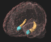 The amygdala (in blue), shown here with the hippocampus, has been linked to a range of mental states such as depression and autism. |
Derived from the Greek for almond, the amygdala sits in the brain's medial temporal lobe, a few inches from either ear. Coursing through the amygdala are nerves connecting it to a number of important brain centers, including the neocortex and visual cortex. "More and more we're beginning to believe, and the evidence is pointing to the idea, that it's the circuits that are important, not just the structure per se," says Ned Kalin, professor of psychiatry, University of Wisconsin-Madison. "And in this particular case the circuitry between the frontal cortical regions of the brain may be critical in regulating emotion and in guiding emotion-related behaviors."
Investigations detailing the amygdala's role date back more than 60 years. H. Kluver and P.C. Bucy reported that amygdala lesions transformed feral rhesus monkeys into tame ones.1 But these lesions were so large and, compared to today's techniques, so crude, that researchers weren't sure of the structures responsible for the behavioral changes. Improved techniques, such as using the neurotoxin ibotenic acid to make more precise lesions; magnetic resonance imaging (MRI); and positron emission tomography (PET) scans of the amygdala's activity, are partly responsible for renewed interest in the amygdala.
A second reason, says John Aggleton, professor of cognitive neuroscience at Cardiff University, Wales, was rat research conducted by Joseph Ledoux, professor of neural science and psychology at New York University, and Michael Davis, professor of psychiatry at Emory University.2,3 This research, says Aggleton, has, in the past 10 years, "paid enormous dividends in understanding how fearful stimuli can control behavior."
But Kalin notes that David Amaral, professor of psychiatry and neuroscience, University of California, Davis, has demonstrated a major difference between rats and monkeys in the links between the amygdala and the rest of the brain.4 "[Amaral's] work has pointed out that there are very strong connections between the amygdala and the neocortex, particularly the visual cortex and the prefrontal cortex. There's not much cross-talk in the rodents between the amygdala and the cortex," says Kalin.
Amaral says he thinks that the amygdala is really playing a protective role. "It is a very phylogenetically old structure," he says. "Probably very early on in Phylogeny, [it] was primarily involved in protecting organisms, moving them away from obnoxious chemical milieu. As organisms evolved it got different kinds of sensory information in to evaluate stimuli in the environment, and that's one of the reasons why it's more highly connected with the neocortex as organisms evolved. It's getting more and more high-level information to do an interpretation of what's going on in the environment."
Kalin's recent monkey study5 shows how a lesioned amygdala can damage that potentially life-saving evaluative function. "We found that when we selectively lesioned the amygdala in 3-year-old animals, the animals' acute fear responses were blunted," he says.
Amaral has found that lesioned adult monkeys abandon their normal caution and tendency to withdraw when confronted with a strange monkey. Instead, they approached the animal and showed "greater frequency and duration of positive social behavior," such as grooming and cooing, than did nonlesioned monkeys.
So striking were these findings, says Bachevalier, that she examined literature on human emotional problems. "When I was starting to read about autism, the behavioral syndrome looked quite similar," she says. "Maybe the amygdala is important in the social deficits we see in this population of humans." She was following up on this hypothesis when Tropical Storm Allison, which hit the Houston area in early June, drowned her study animals.
But Amaral questions whether amygdala lesions in neonatal animals can be used as an autism model. Based on Bachevalier's findings, Amaral lesioned infant monkeys to explore the possible connection,7 and placed them in a cage with an intact monkey. "We expected to see these neonatal monkeys not responding in a social way, like Jocelyne suggested.... We found that the animals did not engage in social interactions, but they are very vigilant and very attentive to the other monkey in the cage. If this was going to be a model of autism, they should show no interest in the other animal. You can get lack of social behavior in a variety of ways. If you're phobic, it's not that you can't read social signals; it's just that you're frightened. That's what our monkeys look like. They're frightened," he says. But Amaral doesn't dismiss the possibility that some amygdala dysfunction could play a role in autism. He is currently using magnetic resonance imaging to examine amygdala function in autistic children and adults.
The overactive amygdala could be a sign of excitotoxicity, a lethal kind of overactivity that kills cells. That, Drevets suggests, might be why a shrunken amygdala is seen in depressed patients. And, he says, some anti-depressive drugs, like lithium, increase biochemical production; these protect neurons from the ravages of overexcitation, thus giving researchers a good reason to continue developing anti-depressants.
While the amygdala is involved in current emotional responses, it is also heavily involved in emotional memory, notes psychology professor Larry Cahill, University of California, Irvine. It gives a "critical boost for long-term memory of emotional events," he says, also noting that men's and women's amygdalas respond differently to emotional situations.
The brain images of women and men were recorded while they were shown emotionally upsetting films, such as plane crashes or killer whales dismembering and eating baby seals. Men showed an increase in glucose metabolism on the amygdala's right side; women showed the increase on the structure's left side. The findings, notes Cahill, don't explain the difference, but they force researchers to explore the basis for it.9 "To my mind, it's saying we have to stop ignoring these kinds of variables [sex differences] when we try to figure out how the brain stores memory for emotional events. Though we only reported the amygdala, in the whole brain we found very different patterns," he says. "We have to now actively incorporate the influence of gender into our theorizing about how the brain stores memories for emotional events, because men and women on average, are probably not doing it in the same way."